Cell signaling is the process of cellular communication within the body driven by cells releasing and receiving hormones and other signaling molecules. As a process, cell signaling refers to a vast network of communication between, and within, each cell of our body. Cell signaling enables coordination within multicellular organisms.

Cell signaling can occur through a number of different pathways, but the overall theme is that the actions of one cell influence the function of another. Cell signaling is needed by multicellular organisms to coordinate a wide variety of functions. Nerve cells must communicate with muscle cells to create movement, immune cells must avoid destroying cells of the body, and cells must organize during the development of a baby.
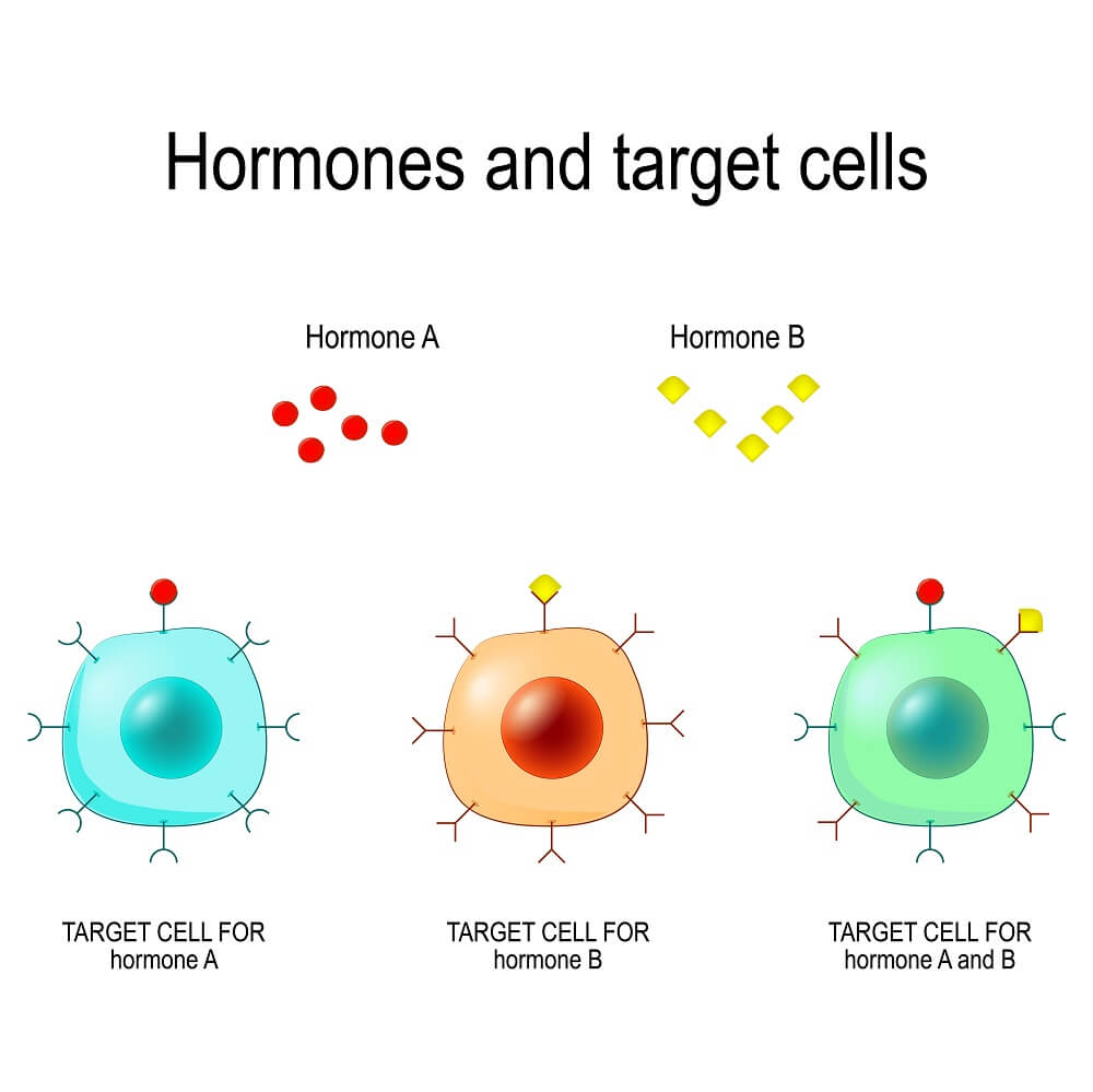
Some forms of cell signaling are intracellular, while others are intercellular. Intracellular signals are produced by the same cell that receives the signal. On the other hand, intercellular signals can travel all throughout the body. This allows certain glands within the body to produce signals which take action on many different tissues across the body. Each target cell will have the required receptors, as in the image below:

Cell signaling is how a tiny gland within the brain can react to external stimuli and coordinate a response. In response to stimuli like light, odors, or touch, the gland can, in turn, release a hormone that activates responses in diverse body systems to coordinate a response to a threat or opportunity.
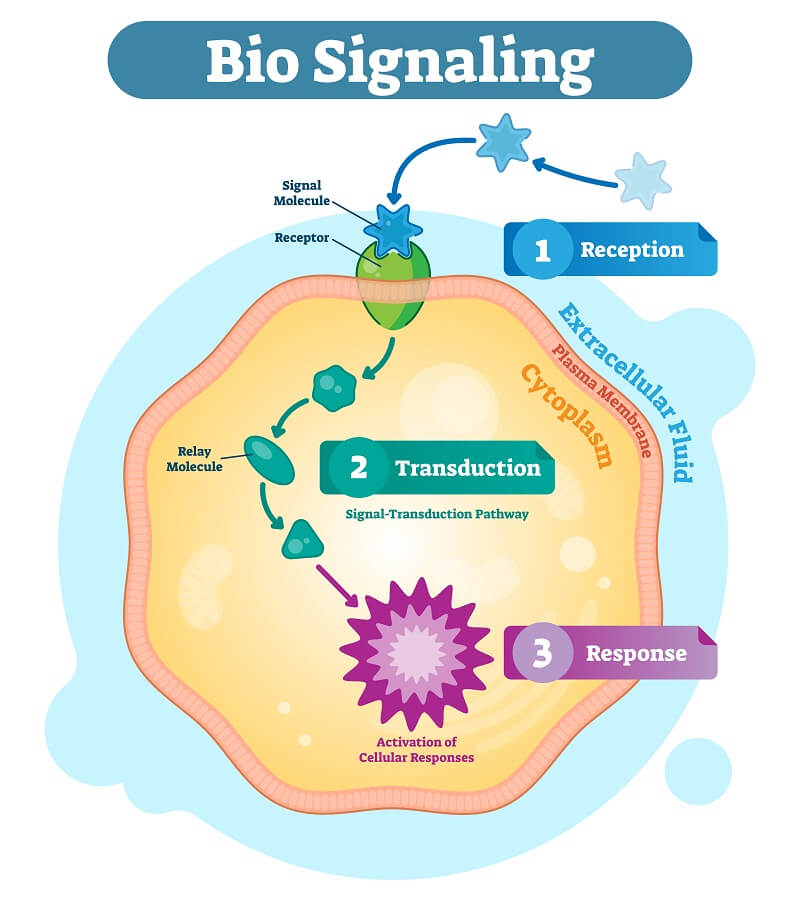
At its core cell signaling can simply be described as the production of a “signal” by one cell. This signal is then received by a “target” cell. In effect, signal transduction is said to have three stages:
Cell signaling serves a vital purpose in allowing our cells to carry out life as we know it. Moreover, thanks to the concerted efforts of our cells via their signaling molecules, our body is able to orchestrate the many complexities that maintain life. These complexities, in effect, demand a diverse collection of receptor-mediated pathways that execute their unique functions.
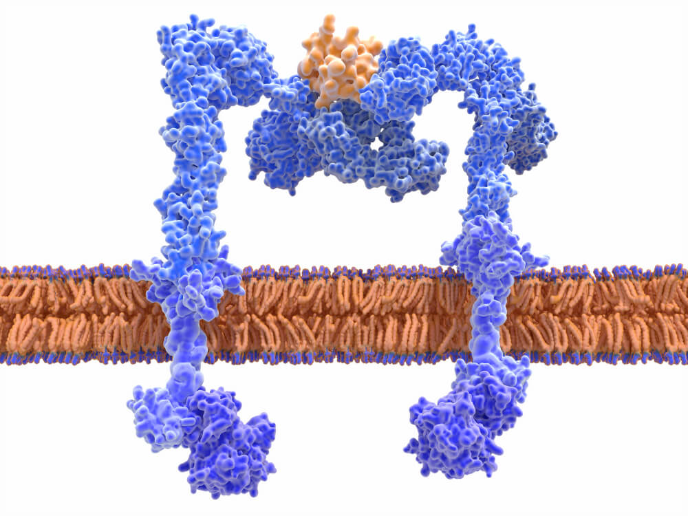
In general, a ligand will activate a receptor and cause a specific response. Receptors are typically protein molecules, as seen in blue below. The orange ligand can be many different types of molecules, but it forms an induced fit with the receptor that is very specific.

A common type of signaling receptor is the intracellular receptor, which is located within the cytoplasm of the cell and generally includes two types. In addition to cytoplasmic receptors, nuclear receptors are a special class of protein with diverse DNA binding domains that when bound to steroid or thyroid hormones form a complex that enters the nucleus and modulates the transcription of a gene. IP3 receptors are another class, which are located in the endoplasmic reticulum and carry out important functions like the release of Ca 2+ that is so crucial for the contraction of our muscles and plasticity of our neural cells.
Spanning our plasma membranes are another type of receptor called Ligand-gated ion channels that allow hydrophilic ions to cross the thick fatty membranes of our cells and organelles. When bound to a neurotransmitter like acetylcholine, ions (commonly K + , Na + , Ca 2+ , or Cl – ) are allowed to flow through the membrane to allow the life-sustaining function of neural firing to take place, among many other functions!
Comparatively, G-protein coupled receptors (GPCRs) remain the largest and most diverse group of membrane receptors in eukaryotes. In fact, they are special in that they receive input from a diverse group of signals ranging from light energy to peptides and sugars. In effect, their mechanism of action also starts with a ligand binding to its receptor. However, the demarcation is that ligand binding results in the activation of a G protein that is then able to transmit an entire cascade of enzyme and second messenger activations that carry out an incredible array of functions like sight, sensation, inflammation, and growth.
Likewise, receptor tyrosine kinases (RTKs) are another class of receptors revealed to show diversity in their actions and mechanisms of activation. For example, the general method of activation follows a ligand binding to the receptor tyrosine kinase, which allows their kinase domains to dimerize. Then, this dimerization invites the phosphorylation of their tyrosine kinase domains that, in turn, allow intracellular proteins to bind the phosphorylated sites and become “active.” An important function of receptor tyrosine kinases is their roles in mediating growth pathways. Of course, the downside of having complex signaling networks lies in the unforeseen ways in which any alteration can produce disease or unregulated growth – cancer. Still, much is yet to be understood about cell signaling pathways, but one appreciable fact is that the importance they carry is nothing short of monumental.
Typically, cell signaling is either mechanical or biochemical and can occur locally. Additionally, categories of cell signaling are determined by the distance a ligand must travel. Likewise, hydrophobic ligands have fatty properties and include steroid hormones and vitamin D3. These molecules are able to diffuse across the target cell’s plasma membrane to bind intracellular receptors inside.
On the other hand, hydrophilic ligands are often amino-acid derived. Instead, these molecules will bind to receptors on the surface of the cell. Comparatively, these polar molecules allow the signal to travel through the aqueous environment of our bodies without assistance.
Signaling molecules are currently assigned one of five classifications.
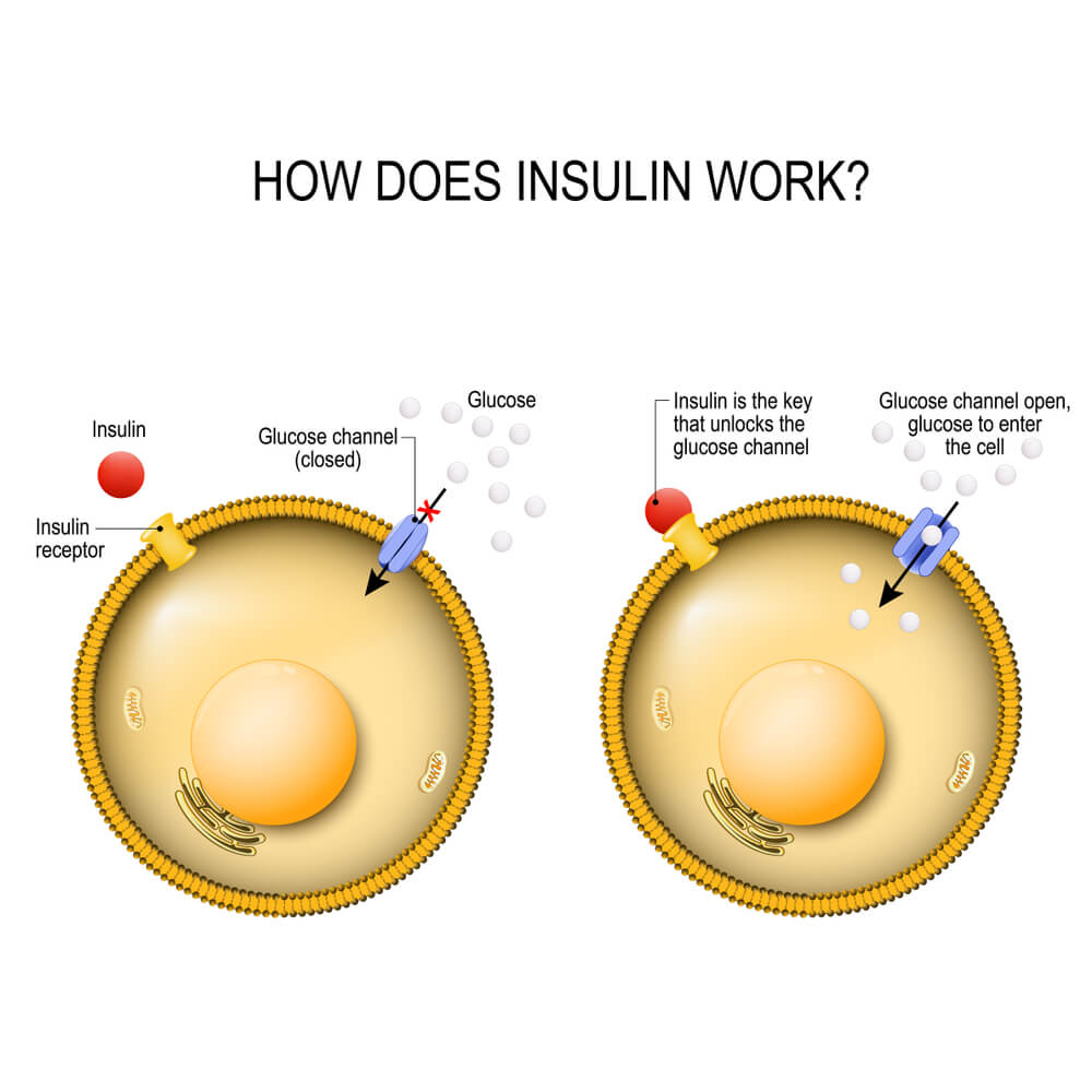
A great (and well-used) example of a cell signaling pathway is seen in the balancing actions of insulin. Insulin, a small protein produced by the pancreas, is released when glucose levels in the blood get far too high.
First, the high glucose levels in the pancreas stimulate the release of insulin into the bloodstream. Insulin finds its way to the cells of the body, where it attaches to the insulin receptors. This sets off a signal transduction pathway within each cell that causes the glucose channels to open, as seen in this graphic:

As glucose flows into the cell, the glucose levels in the bloodstream are slowly decreased. The cells will use the glucose to create ATP energy or the cells store it as fats and starches for later use. Once the glucose level in the bloodstream has dropped to a sufficient level, the pancreas stops producing insulin, and the cells shut down their glucose channels.
Bruice, P. Y. (2011). Organic chemistry (6th ed). Boston: Prentice Hall.
Lehninger, A. L., Nelson, D. L., & Cox, M. M. (2008). Lehninger principles of biochemistry (5th ed). New York: W.H. Freeman.
Lodish, H. F. (Ed.). (2008). Molecular cell biology (6th ed). New York: W.H. Freeman.